Pressure Injuries That Start in the Operating Room: What the Surgical Team Needs to Know

Pressure injuries can begin in the operating room long before anyone sees them. Across 35 studies, a 2025 systematic review reported a mean incidence of 17.22% among surgical patients, or roughly one in six (1).
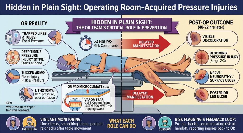
Picture a six hour robotic pelvic procedure that goes well. The patient is extubated, stable, and arrives in PACU with skin that looks normal. Two days later, on the step down unit, a deep purple area appears over the sacrum. It is charted as a unit event, and nobody in the OR hears about it.
The surgical team may never see the injury it helped start. Tissue damage can begin while the patient is anesthetized and immobile, then become visible only after the patient reaches recovery or the nursing unit. Positioning, support surfaces, padding, warming, case duration, and perfusion are all set in the OR, so prevention belongs to everyone in the room.
How common is it?
Published rates vary with population, procedure, definition, and follow up, but systematic reviews consistently show a clinically meaningful incidence.
Population | Reported rate | Source |
All surgical patients, 35 studies | 17.22% mean incidence | (1) |
All surgical patients, 17 studies | 15% pooled incidence | (2) |
Cardiac surgery | 18% pooled incidence | (3) |
Open heart surgery | 24.06% pooled prevalence | (4) |
Spine surgery | 23.0% incidence | (5) |
Mixed surgical populations, single hospitals | 5.8% to 8.4% |
The 2025 review found the highest incidence in orthopedic studies, particularly hip surgery, and noted that a 2012 review reported about 15% (1). Similar pooled rates more than a decade apart mark this as an unresolved perioperative problem.
Some literature attributes up to 45% of hospital acquired pressure injuries to surgery (8). That is an upper estimate. Other reports put the share near 23% for procedures longer than three hours (9). The exact percentage varies. The point does not: a meaningful share of pressure injuries begin during surgical care.
The injury may appear after the OR team has moved on
Skin can look normal when the procedure ends even though damage has begun. Studies commonly evaluate operating room acquired injuries that develop within 48 to 72 hours of surgery (6). Most appear one to three days after the procedure, and some as late as day six (5).
- A normal PACU skin assessment does not clear the case. Tissue exposed to prolonged pressure, deformation, heat, moisture, or shear may not show surface injury right away.
- The receiving unit may not associate the wound with surgery. By the time discoloration, blistering, or necrosis appears, the patient has been out of the OR for days (10).
- The surgical team may never get feedback. If the injury is documented only as a nursing unit event, the clinicians who chose the position, surface, padding, and warming never learn the outcome.
Without that feedback loop, the same conditions get repeated.
Who is at highest risk?
Time on the table is one of the most consistent risk factors, and it compounds with position and patient condition.
Duration. Prior research cited by Kirkland‐Walsh and colleagues reported that after four hours of surgery, risk rose about 33% for each additional 30 minutes (11). In one U.S. population of 1,128 patients, the rate rose from 5.8% in procedures under 180 minutes to 34.8% in procedures over 480 minutes (12). Duration is cumulative tissue exposure, not only operative time.
Position. Steep Trendelenburg for robotic pelvic procedures places patients about 30 to 40 degrees head down (15). Risk of nerve injury, pressure injury, and deep tissue ischemia rises with the angle and the time in position (14). One operating room study identified prone positioning as a significant risk factor, with an odds ratio of 3.35 (13).
Tucked arms. Arm tucking is common in robotic and lithotomy cases, and it carries a measured nerve injury risk. In 324,124 anesthetics, persistent ulnar neuropathy occurred in 6.8 per 100,000. The absolute risk is low, but tucked arms were associated with 6.16 times greater odds (95% CI 1.85 to 20.59), and the odds also rose with BMI and with each added hour of surgery (26). Across robotic procedures in lithotomy with steep Trendelenburg, reported nerve injury rates range from 0.16% to 10.0% (27). These are nerve injuries, not pressure injuries, and the incidence of hand, wrist, and arm pressure injury with tucked arms has not been well quantified. The same forces are at work, and tucked arms are hard to inspect once the patient is draped and the robot is docked.
Compartment syndrome of the hand and forearm has also been reported with tucked arms. The evidence is case reports, and no incidence has been published. One report and literature review described hand compartment syndrome requiring emergency fasciotomy after a prolonged robotic rectal resection in Trendelenburg. It named high BMI, arm tucking, and long duration as the shared risk factors, and noted earlier cases caused by draw sheets tucked too tightly around the arms (29).
Patient factors. Age, hemodynamic status, albumin level, blood loss, and anesthesia related factors have all been associated with injury (6, 7). Diabetes carries about 1.74 times greater odds overall and 2.00 times among cardiac surgery patients (17).
The highest risk patient is often identifiable before incision: a long procedure, a demanding position, compromised physiology, and no ability to move. That should trigger prevention before positioning, not after skin changes appear.
Where equipment causes harm
Not every pressure injury comes from the table mattress. In a cohort of 403 surgical patients, 23.5% of those who developed injuries had injuries associated with positioning devices, and 11.8% had injuries associated with devices or instruments (7). In another cohort, 4.2% of patients with medical devices in place developed a device acquired injury (6).
- Devices and instruments: airway devices and ties, masks, blood pressure cuffs, oximeter probes, monitoring cables, lines, stopcocks, grounding pads, tourniquets, and instruments resting on the body.
- Positioning equipment: headrests, prone head supports, arm boards, stirrups, leg holders, bolsters, lateral supports, bean bags, safety straps, traction boots, and perineal posts.
Lithotomy stirrups. In a 2026 prospective study of 218 urologic patients in lithotomy, 57 patients (26.1%) had 114 stage 1 pressure injuries at the end of surgery, and every injury was on the posterior legs (25). No injury was detected at the 24, 48, or 72 hour checks, so these were early, reversible changes and not lasting wounds. They still show where the load goes. The weight of the leg rests on a narrow area of calf and heel, elevation lowers perfusion pressure to that same tissue, and the legs stay fixed while the torso can shift when the table tilts. The authors recommend supporting the posterior legs.
Compartment syndrome in the legs. The same position can cause well leg compartment syndrome, in which rising pressure inside the calf compartments cuts off perfusion to muscle and nerve. Reported incidence is about 1 in 3,500 abdominopelvic procedures in lithotomy, and as high as 1 in 500 after cystectomy (30). Across 3,110 robotic prostatectomies at 17 UK centers, nine patients developed it (0.29%) and seven needed fasciotomy. Eight of the nine had console time over four hours and five were obese (31). The risk factors match the rest of this article: time, head down tilt, calf compression, and body mass.
Anything hard, narrow, folded, wrinkled, or trapped between the patient and the surface concentrates load on a small area for the whole procedure.
What the table surface does and does not do
A pressure redistributing surface can reduce interface pressure. It cannot make a long case safe on its own. In a randomized comparison, viscoelastic foam produced lower average interface pressure than gel or a standard table in the supine position, with no difference in prone (20). How the surface behaves clinically also depends on thickness, temperature, patient weight, position, moisture, warming, shear, and duration.
Viscoelastic foam is temperature sensitive by design. Memory foam becomes firmer as temperature drops and softer and more compliant as it warms, and its firmness, support, and recovery also change with humidity (19). In a cool operating room the pad starts firm. Once the patient is positioned, body heat, draping, moisture, and active warming change the surface through the case. The temperature that matters is at the patient and surface interface, not in the room.
The size of the change has been measured. In patent testing by a foam manufacturer, twelve viscoelastic foams lost 15% to 54% of their firmness when warmed from 72°F to 100°F. The same patent reports a commercial viscoelastic foam measuring 35 pounds of indentation force at 50°F against 4 pounds at 70°F (28). This is a property of the material, not a hypothesis.
Not all foams respond this way. The same patent notes that typical polyurethane foam passes through its glass transition near −50°C, far below any clinical temperature, while viscoelastic foam is formulated to make that transition close to room temperature (28). A conventional foam therefore changes relatively little between a cool room and a warmed patient. A viscoelastic foam spends the whole case inside its transition range, which is why its firmness moves so much.
Thickness matters. Many positioning systems use thin foam overlays. A thin layer has less depth available before it is fully compressed, and as the foam warms under sustained load that margin can shrink further (24). Some envelopment is desirable because it spreads load. Bottoming out is not, because the surface underneath then determines what the tissue experiences. The question to ask is whether the complete support system keeps redistributing load under this patient’s weight, at clinical temperature, for the expected duration.
Warming changes the result. In a randomized cardiac surgery trial, patients on a 4 cm viscoelastic foam overlay over a water filled warming mattress developed pressure ulcers at 17.6%, compared with about 11% on the warming mattress alone. The investigators concluded the combination could not be recommended (18). The trial does not show that every viscoelastic surface or warming system raises risk. It suggests that a material that redistributes pressure in isolation may not deliver the same result inside a loaded, layered, and warming system.
The draw sheet is part of the surface. A draw sheet lies between the patient and the pad for the entire case. Laboratory testing on bed surfaces found that added linen layers significantly reduced the surface’s ability to withdraw heat and evaporate moisture, and plastic backed layers reduced it most (22). A review of microclimate testing reached the same conclusion for linen layers and drawsheets (23). Those tests were done on low air loss bed surfaces, not operating tables. The principle carries over: an absorbent sheet over a pad with a fluid proof cover restricts moisture vapor transfer and becomes the place where moisture collects. In effect it acts as a sump against the skin. A wrinkled or bunched sheet also adds focal pressure and shear.
Moisture needs a way out. The surgical patient does not move. An awake patient shifts, and each shift lets air reach the skin and moisture escape. Under anesthesia the same skin stays pressed against the same surface for hours, so the only route out for heat and moisture is through the surface itself.
OR surfaces are built as fluid barriers. Gel pads and table pads sealed in polyurethane or vinyl skins are made impermeable on purpose, for infection control and cleaning between cases. They are chosen because they are reusable and easy to clean, and they are often seen as the responsible alternative to disposable foam. The barrier that keeps fluid out also keeps vapor in. These hydrophobic surfaces do not absorb sweat and pass little or no vapor, so moisture stays at the skin for the length of the case, and damp skin tolerates pressure and shear less well (21). Layers that restrict heat and vapor transfer have been shown in surface testing to hold moisture at the interface (22, 23).
Inpatient surfaces made a different choice. Hospital mattresses face the same cleaning demands, and they commonly use vapor permeable covers that block liquid while letting moisture vapor leave. Vapor transmission is a routine part of how those surfaces are evaluated. For OR table surfaces, published vapor transmission data are scarce.
The cost was never part of the decision. Reusability and cleanability drove the choice of OR surfaces. The physiological cost of an impermeable surface under a patient who cannot move was not part of that discussion. Absorbent, vapor permeable layers can close the gap. Until the data exist, ask each vendor for the vapor transmission rate of the surface that touches the patient.
Pressure is only part of the problem. Temperature and humidity at the skin affect its structure and its tolerance for pressure and shear (21). In laboratory studies of healthy volunteers, a 1°C rise in skin temperature contributed as much to skin ischemia as roughly 8 to 14 mmHg of added interface pressure, and the effect was strongest above 32°C (32, 33). Clinical evidence from the operating room points the same way. In a prospective study of 29 patients having surgery in the park bench position, seven (24.1%) developed pressure ulcers, and skin microclimate measured continuously through surgery was an independent risk factor (34). The number of clinical studies is still small, which is a reason to measure microclimate in the OR and not a reason to discount it. Pressure mapping at room temperature cannot describe what is happening under an anesthetized patient several hours into a warmed procedure. The complete system matters.
What each role can do
The evidence points to the same interacting factors: time, position, equipment, support surfaces, perfusion, temperature, moisture, and delayed detection. Apply these practice considerations within institutional policy, manufacturer instructions for use, and current professional guidance.
Before the case, as a team
- Flag patients with an expected procedure over three hours, extreme or prolonged positioning, advanced age, diabetes, impaired perfusion, or poor nutritional status.
- Inspect and document skin over the sacrum, heels, occiput, and position specific pressure points.
- Discuss the planned position and support surfaces before positioning begins.
- Know every layer under the patient: table mattress, positioning devices, warming equipment, pads, sheets, and grounding equipment. Every layer changes how the system handles load, heat, and moisture. Ask what each one does with sweat and vapor, not only with pressure.
Circulating nurse and scrub team
- Smooth every layer beneath the patient. Remove folds, wrinkles, and unnecessary materials, including extra sheets.
- Verify that lines, cables, stopcocks, and connectors are not trapped beneath the body.
- Inspect contact points at arm boards, stirrups, headrests, straps, and lateral supports.
- Keep instruments and team members from resting on the patient.
- Recheck the patient after every significant table movement or position change.
Arms and legs in lithotomy and robotic cases
- When arms are tucked, keep the forearms neutral, pad the elbows and hands, and confirm that fingers are clear of table joints and that lines are not pressing into skin. Do not wrap the tucking sheet tightly around the forearm or hand.
- Inspect tucked arms and hands before draping and docking, because access is limited afterward.
- In stirrups, spread the load along the whole calf, keep the heel seated, and keep pressure off the outer knee.
- Recheck legs and arms after every tilt, and keep people and equipment from leaning on raised legs.
Anesthesia
- Periodically inspect the occiput, ears, face, and airway device contact points, particularly in prone and steep head down cases.
- Keep tube ties, masks, cuffs, probes, and monitoring equipment from exerting continuous focal pressure.
- Treat sustained hypotension as a tissue perfusion problem as well as a hemodynamic event.
- Together with the circulating nurse, call out the three and four hour marks so duration stays in the team’s awareness.
Surgeon
- Use the least extreme position that gives adequate exposure. One study found benign robotic gynecologic surgery was performed effectively at a mean of 16.4 degrees of Trendelenburg (16).
- Support periodic position and pressure point reassessment in long procedures, and relief from extreme positions when clinically feasible.
- Treat operative time as a patient exposure variable, not only an efficiency metric.
Handoff and the receiving unit
- Communicate the position, duration, major positioning devices, areas of concern, and any abnormal skin findings.
- For high risk cases, tell the receiving unit that a normal immediate exam does not exclude developing injury, and that skin checks should continue through 72 hours.
- Report suspected intraoperatively acquired injuries back to the OR. The team that positioned the patient needs to know the outcome. Make this a standing report from the units to the OR, because without it the team cannot refine its positioning protocols or judge how its surfaces perform.
The anesthetized patient cannot feel discomfort, shift weight, or report a cable underneath them. A pressure redistributing surface is one component of prevention, not a substitute for it. The safest approach treats the patient and the whole positioning environment as one system, from induction through postoperative surveillance.
Sources
- Kurian, Moore, Patton, George. The Incidence of Pressure Ulcers in Surgical Patients: A Systematic Review. International Wound Journal, 2025.
- Chen, Chen, Wu. The Incidence of Pressure Ulcers in Surgical Patients of the Last 5 Years. Wounds, 2012.
- Chen et al. Surgery related pressure ulcers in cardiac surgery patients. International Wound Journal.
- Prevalence of pressure injury in open heart surgery: systematic review and meta analysis.
- Li et al. Prediction model for intraoperatively acquired pressure injuries in orthopedics. Frontiers in Physiology, 2023.
- Savcı, Karacabay, Aydın. Incidence and Risk Factors of Operating Room Acquired Pressure Injury, 2024.
- Ozdemir et al. Pressure injuries in the operating room: who are at risk? Journal of Wound Care, 2023.
- Fazzalari et al. Mitigating perioperative pressure injuries in microsurgical breast reconstruction. Frontiers in Surgery, 2025.
- Preventing pressure injuries in the operating room. American Nurse Today, 2018.
- Intraoperatively Acquired Pressure Ulcers: Are There Common Risk Factors? Wound Management and Prevention, 2007.
- Kirkland‐Walsh et al. Pressure Mapping Comparison of Four OR Surfaces. AORN Journal, 2015.
- Length of surgery and pressure ulcers risk in cardiovascular surgical patients: a dose response meta analysis.
- Evaluation of pressure ulcer risk and development in operating rooms, 2022.
- Steep Trendelenburg support surface trial registration.
- Trendelenburg position reference. STERIS.
- Ghomi et al. Trendelenburg position in gynecologic robotic assisted surgery.
- The Association between Pre-existing Diabetes Mellitus and Pressure Ulcers in Patients Following Surgery: A Meta-analysis. Scientific Reports.
- Feuchtinger et al. A 4 cm thermoactive viscoelastic foam pad on the operating table to prevent pressure ulcer during cardiac surgery, 2006.
- Viscoelastic (Memory) Foam. Polyurethane Foam Association, InTouch.
- Comparison of interface pressures on three operating table support surfaces during surgery.
- Microclimate: A critical review in the context of pressure ulcer prevention. Clinical Biomechanics, 2018.
- A laboratory study examining the impact of linen use on low air loss support surface heat and water vapor transmission rates, 2013.
- Clinical Relevance of Microclimate Tests on Support Surfaces. Advances in Skin and Wound Care.
- The Warming Foam Problem. Author’s supporting material, not peer reviewed. Used for the discussion of foam thickness and thermal response.
- Karaca, Okgün Alcan. Lithotomy Position and Intraoperative-acquired Pressure Injury: A Prospective Observational Study. Advances in Skin and Wound Care, 2026.
- Pulos et al. Perioperative Ulnar Neuropathy: A Contemporary Estimate of Incidence and Risk Factors. Anesthesia and Analgesia, 2021.
- Intraoperative peripheral nerve injury related to lithotomy positioning with steep Trendelenburg in robotic assisted laparoscopic surgery: a systematic review.
- Tursi, Chan, Niederoest. Low energy-loss, high firmness, temperature sensitive polyurethane foams. US Patent 6,653,363, Foamex L.P., 2003.
- Hand Compartment Syndrome After Prolonged Robotic-Assisted Laparoscopic Rectal Resection: A Case Report and Literature Review.
- Avoiding, diagnosing and treating well leg compartment syndrome after pelvic surgery. British Journal of Surgery.
- Pridgeon, Bishop, Adshead. Lower limb compartment syndrome as a complication of robot-assisted radical prostatectomy: the UK experience. BJU International, 2013.
- Lachenbruch, Tzen, Brienza, Karg, Lachenbruch. The Relative Contributions of Interface Pressure, Shear Stress, and Temperature on Tissue Ischemia: a Cross-sectional Pilot Study. Ostomy Wound Management, 2013.
- Lachenbruch et al. Relative Contributions of Interface Pressure, Shear Stress, and Temperature on Ischemic-induced, Skin-reactive Hyperemia in Healthy Volunteers. Ostomy Wound Management, 2015.
- Yoshimura, Nakagami, Iizaka, et al. Microclimate is an independent risk factor for the development of intraoperatively acquired pressure ulcers in the park-bench position: a prospective observational study. Wound Repair and Regeneration, 2015;23(6):939-947.
Figures and clinical statements are drawn from the cited literature and supporting material. This article is for team education and does not replace facility policy, manufacturer instructions for use, or clinical judgment.
Related Posts

The Infinitus Advantage
Please take just five minutes to discover how we stand apart—and how your legacy surgical…

Here is a Sneak Peek of Our Upcoming Pneuma® Genesis Infinity Platform!
https://vimeo.com/1127575396?fl=pl&fe=sh Please reach out to your local surgical care representative, or contact us at infinitusmedical.com…

Here Is a Sneak Peek of Our New Luna SP®
Come see how our newest supine comfort pad can be accessorized for your surgical procedural…